In the third installment dedicated to research in Brain related areas, we chat to Professor Mario Valentino about the Laboratory for the Study of Neurological Disorders’s latest finds, and about how these are ushering in a new era in real-time visualisation of the dynamics in stroke onset for the treatment of strokes.
 In 2012, the European Society of Cardiology and the European Heart Network released statistics that placed stroke at number two on the list of the single most common causes of death in Europe. In fact, some 1,100,000 people die from it annually on our continent alone.
But what is a stroke exactly?
“The brain is the most complex system in the universe consisting of 100 billion neurons which are interconnected in a still mysterious way,” explains Professor Mario Valentino, who, upon his return to Malta from stints at the Max Planck Institute for Neurological Research in Germany, and the Department for Neurology at Washington University and the Hope Centre for Neurological Disorders in the USA, set up the Laboratory for the Study of Neurological Disorders here at the University of Malta.
“Electrical signals coursing through this awesome network somehow underlie our perception of the world and our actions within it,” he continues. “Because of all this, the brain is an energy-hungry organ, and despite comprising of only 2% of the body’s weight, the brain consumes more than 20% of our daily energy intake.
When the blood supply to the brain is interrupted or blocked for any reason, the consequences are usually dramatic. Control over movement, perception, speech, and other mental or bodily functions can be impaired, and consciousness itself may be lost. Deterioration continues over hours, or even days, and depends primarily on the severity and the duration of the ischaemia [inadequate blood supply to the brain].
“Despite major advances in prevention and rehabilitation, few neurological injuries are as debilitating as stroke. The disease is currently the third leading cause of death after heart disease and cancer and the leading cause of long-term disability worldwide; it is similarly devastating in Malta.”
What’s worse is that, the already-staggering numbers are expected to grow in the years to
come. This is mostly due to the enhanced susceptibility to stroke at an older age as a result of an increase in life expectancy.
“The magnitude of the problem on pre-term babies is equally extraordinary and the main cause of cerebral palsy, the most common neurological disorder of infancy,” Professor Valentino adds. “Unfortunately, current options for acute treatment are extremely limited and there is an urgent need for new treatment strategies. After all, fast and timely restoration of blood flow is imperative for recovery after a stroke.”
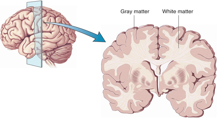
Over the last few years, Professor Valentino, along with the members of the Laboratory for the Study of Neurological Disorders, has been working on understanding how white matter (matter found in the brain that actively affects the way the brain learns and functions) could actually play a part in strokes.
“Stroke was once considered a disorder of blood vessels, yet growing evidence has led to the realisation that the biological processes underlying stroke are driven by the interaction of neurons, glia [supporting cells of the nervous system], vascular cells and matrix components, which actively participate in the mechanisms of tissue injury and repair,” he says.
“That’s why we have to understand how the injuries are occurring if we are to prevent them from happening, and that’s what driving us to study cell-to-cell interactions in the normal and diseased nervous system.”
For five years, the team has sought to understand not just the properties of individual cells, but also the dynamics of the loss of their interactions during a stroke. To do this, they have set-up a powerful technique for in vivo (occurring in a living organism) imaging that is centred around two-photon microscopy (a fluorescent imaging system that combines the use of a pulsed infra-red laser) that allows them to visualize in real-time the cellular workings of the brain.
“The mastering of two-photon microscopy combined with the use of mouse models whose different cell populations shine in different colors allow for simultaneous multicolor imaging, as well as unlimited number of examinations of the same field of view through a small ‘window’ in the skull. This opens the way to quantitative and correlative analysis of cell distributions and interactions in a truly physiological environment.
“In addition, large amounts of data are collected from a single animal, hence allowing for a reduction in the number of animals engaged in an experimental study and improving the significance of the results.”
As part of this study, Professor Valentino and his team have recently shown that the vulnerability of white matter to stroke varies during development, and that the metabolism of the brain’s wiring revolves around lactate that is provided by  astrocytes, the main supporting cells in the nervous system.”
It is clear that up to 30% of all strokes in humans occur as infarcts deep in the white matter – so much so, that these are what produce specific stroke impairments, including a weakness on one side of the body and cognitive decline. Moreover, white matter has been linked to many other disorders, including dementia and several leucodystrophies in children.
“While the pathophysiology of grey matter [a main component of the central nervous system] in stroke has been intensively studied, much less attention has been focused on the white matter, which comprises half of the volume of the human brain,” says Professor Valentino. “Most fibers in white matter serve to connect local or contralateral brain areas, and there is growing recognition of the potential neurocognitive consequences of white matter lesions.
“It is important to point out that the basic research in this area has been limited because of the difficulty in establishing an animal model of subcortical white matter stroke and the minimal volume of white matter volume in rodents,” he continues. “Yet, since neuronal death is seen as a consequence of the failure of astrocytes to support the metabolic demand of neurons, we have proposed that new efforts should be designed to protect the integrity of astrocytes as an alternative strategy for neuroprotection.” This perspective is centred around their recent finding that astrocytes are more vulnerable to ischaemic injury than previously thought.
All this, Professor Mario Valentino hopes, will develop better stroke therapies and help scientists, researchers and doctors understand how the brain’s vasculature (vascular system), and its interaction with neurons and glia cells, is altered after a stroke.
“It is this knowledge of the intricate dynamics and cross-talk between neurons, glia and the vasculature that is unraveling what causes these cells to die, and which might one day lead us to understand how they could be made to recover,” Professor Valentino concludes.
In other words, this study currently being undertaken at the University of Malta is trying to understand what goes wrong in the brain before, during and after someone experiences a stroke, in the hope of being able to rectify the short- and long-term consequences it has on the patient’s brain and life. And while we may not know the outcome for another couple of years, it’s definitely a very promising start.
You too can be part of this fascinating world of research by supporting researchers in all the faculties of the University of Malta. Please click here for more information on how to donate to research through the Research Trust (RIDT).
